Clinical Case Reports
Subharti Dental College & Hospital, Meerut
Interceptive orthodontics, space preservation therapies, dentofacial orthopedics, clear aligners, and comprehensive case reports from the Department of Orthodontics.
Case Report 1
Orthodontics & Dentofacial Orthopedics

Case Report 2
Orthodontics & Dentofacial Orthopedics
Case Report 3: Sequential Utilization of “e” Space
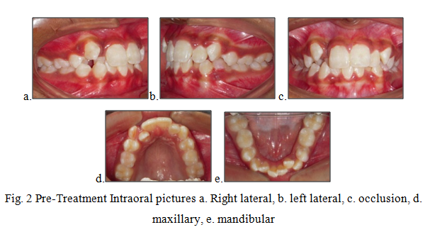
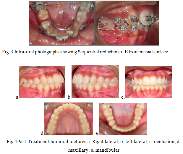
Mixed Dentition OrthodonticsLeeway space and E-space plays a crucial role in the late transition phase from primary dentition to permanent dentition. A 9-year-old female patient reported with a chief complaint of irregularly placed upper and lower front teeth. On extraoral examination (Fig. 1a.,b.c.), she had a straight profile, unesthetic smile with a prominent maxillary canine on the right side. Intraoral examination (Fig. 2 a.-e.) revealed that she was in the late mixed dentition stage with Angle’s class I molar relation on both sides. Maxillary midline was shifted to the right by 1.5 mm with respect tofacial midline, and mandibular midline was shifted to left by 1 mm. All deciduous second molars (E’s) were present. Maxillary right lateral incisor was in crossbite. Based on her dental age and model and cephalometric analysis, it was decided to use E-space for correction of crowding in anterior teeth and guided eruption of permanent second premolars. Nance’s palatal arch and lingual holding arch were constructed and cemented in upper and lower arches, respectively. Arch alignment was started with partial bonding in both arches. E’s were sequentially reduced at the rate of 1 mm/month from the mesial side for distalization of first premolars and canines with tie backs in all four quadrants. Alignment was improved significantly in both arches till the time of shedding of E’s (Fig. 3). By the end of the treatment, both the arches were aligned completely without any significant proclination of incisors. Molars were maintained in class I relationship, and canines were brought into class I relationship. There was perfect alignment as seen in occlusal photographs (Fig. 4). Profile of the patient was satisfactory after treatment (Fig. 5).