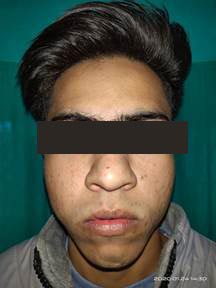
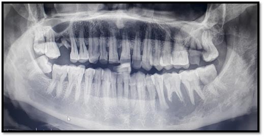
18 years old male patient reported with a chief complain of pain in right upper tooth region since 15 days. The patient had pain and discomfort in eating food and mild swelling was seen on right side of face.
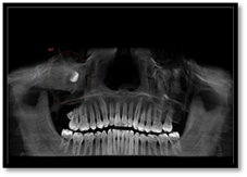
OPG revealed the presence of impacted maxillary third molar on right side
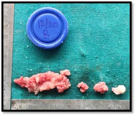
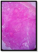
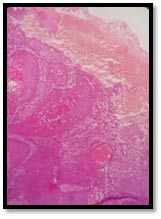
Multiple bits of soft tissue specimen were received for histopathological confirmatory diagnosis.
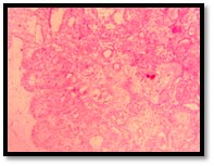
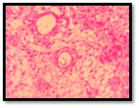
Histopathological examination revealed presence of ameloblast like cells arranged in network enclosing central stellate reticulum like cells. Few areas showed pseudoglandular arrangement of ameloblast like cells. Ghost cells were also evident. The final diagnosis was
Plexiform Ameloblastoma.
The patient was treated with wide surgical enucleation.
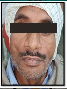
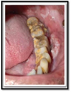
A 32 year old male patient reported with painful ulcer in left lower back tooth region since 1 month and had difficulty in chewing on left side. Patient had poor oral hygiene and he gave the history of smoking bidi and chewing gutkha. Intraorally an ulceroproliferative, exophytic growth was seen in lower left buccal vestibule extending from 34 to 37
OPG revealed that the radiolucency extending from left second premolar to second molar and root resorption was also evident.
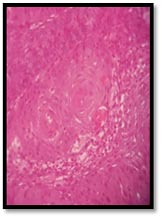
4x 10x 40x
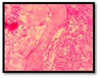
4x-- H &E stained section shows connective tissue stroma being invaded by numerous dysplastic epithelial islands.
10x-- H &E stained section shows well differentiated dysplastic island epithelial with moderate amount of keratin pearl formation.
40 x—Few dysplastic features like cell pleomorphism , hyperchromatism, altered N/C ratio are seen and inflammatory cells are present in moderate amount. Final Diagnosis of Well Differentiated Oral Squamous Cell Carcinoma was rendered. Patient underwent radical neck dissection and was further referred for Radiation Therapy.
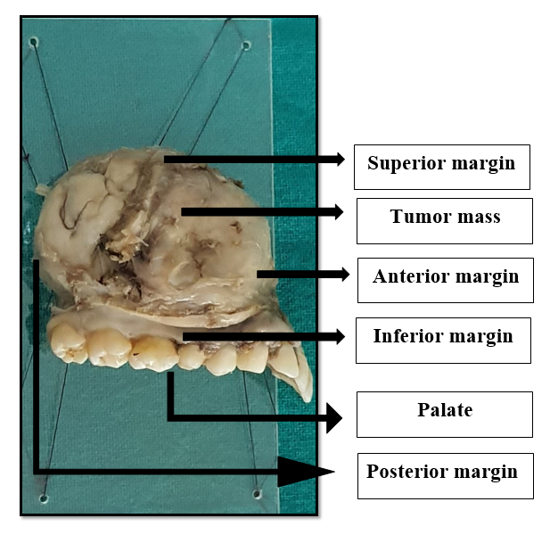
Rare case of Juvenile Ossifying Fibroma reported to Department of Oral Pathology & Microbiology
History of patient: A female patient aged 19 years reported to Subharti Dental College & Hospital, Meerut; with a chief complaint of swelling over right side of face since 6 months.
Provisional diagnosis: Juvenile Ossifying Fibroma
Gross specimen:
Histopathological diagnosis:
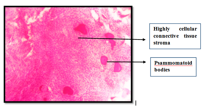
H & E stained section shows extremely fibro cellular connective tissue stroma showing predominately plump fibroblasts and few collagen fibres. Numerous eosinophilic ossifications are seen within the stroma. Concentric lamellated calcified structures with brush borders resembling psammoma bodies are also seen. Bony trabeculae are also evident.
Above mentioned features were suggestive of Juvenile Ossifying Fibroma (Psammomatoid variant)